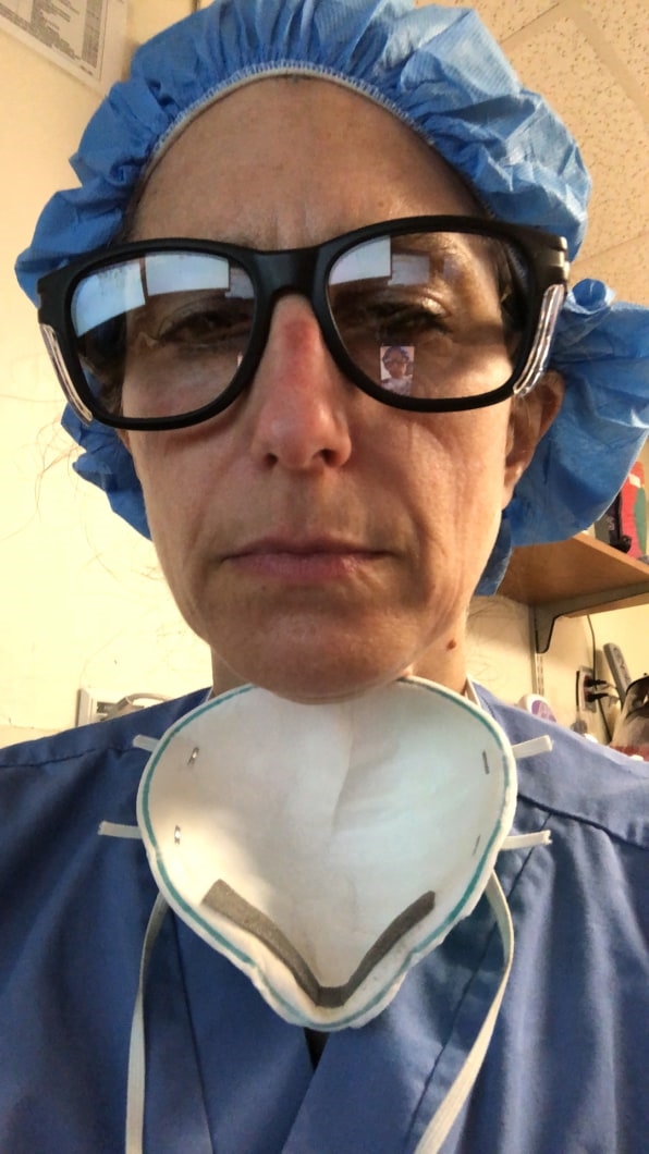
Tall and thin, wearing blue jeans, the patient walked past me in the emergency department. Wearing a surgical mask, he walked behind the triage nurse following her to the patient room and got hooked up to the heart rate, blood pressure, and oxygen monitor. When I entered, rolling a cart with an ultrasound machine the size of a laptop, I heard the fast beeping accompanying the rate of his heartbeat. I was wearing the uniform that’s become standard in the last two months: the awkward set of equipment and garments that people have come to know as PPE (personal protective equipment). Bouffant hat, light-blue plastic gown wrapped and tied at the waist, oversized goggles, an aqua-green N-95 mask which was driving me nuts by chafing against the bridge of my nose, and purple gloves—size small.
He was one of my first COVID-19 patients. He had a fever and a normal oxygen level, telling me that he’d been sick with no appetite and just tired and weak. He seemed unaware of how fast he was breathing and yet he had a scared expression on his face the times I entered the room. Since the gown opens in the back, I could see how quickly he was gasping for air. When I placed the ultrasound probe on the right side of his back, just below the shoulder blades, I saw something familiar on the screen. It was the now well-known picture that my colleagues in China and Italy had been describing to us here in the U.S. for months: a black-and-white snowstorm of viral infection in the lungs.

[Photo: courtesy of the subject]
I work at Thomas Jefferson University Hospital, one of Philadelphia’s largest healthcare facilities where, unlike New York City emergency departments, the pace is still quiet with episodic surges and steadily increasing volume. We have a stable supply of PPE, though it’s rationed. And our ICU beds are almost full of COVID+ patients. But in anticipation of the storm to come, we’re feeling anxious. The rules and the numbers change daily and are climbing weekly. Right now, the emergency department patient visits are down. So if people come in, they are sick. It was the middle of my shift, and one of my first days putting on and taking off PPE. There was a lot rolled into this first experience a month ago now, all centered around maintaining deliberate focus: to care for and communicate with patients, check and confirm COVID-19 patient care algorithm, stabilize them, reassure them. And deliberate focus reflected inward: don’t touch your face, take off part of the PPE in the room, and hand hygiene. Hand hygiene. Did I mention hand hygiene?
And we’re learning some key ways to hasten the diagnosis and treatment of all these patients, including the adaptation of technology that we’ve been using for decades. Ultrasound gave me a clue to his condition long before a nasal swab would deliver results. Those take hours to days depending upon the laboratory. Not every patient needs an ultrasound or should get an ultrasound test. But some patients do benefit, and by working so quickly to inform us about a patient’s condition, it allows us to hasten their treatment.
Simply put, ultrasound works by electricity causing crystals to vibrate. The vibrations emit sound waves through a liquid medium (in this case, ultrasound gel). The ultrasound waves enter the body, hit an object of interest—such as lung tissue—and reflect back to the probe. The reflected waves are translated onto the screen. Ultrasound performed at the point-of-care is broken down into three parts: the clinician (often a doctor) performs the ultrasound beside the patient, then interprets the ultrasound images on the screen in real time, and then integrates the findings into the patient’s care. Ultrasound just may play a pivotal role and be the understated technology solution for doctors triaging, diagnosing, and following the progress of the COVID-19 disease. Right now we are not sure which subset of patients benefits most. And we are not sure when in the algorithm ultrasound should enter the decision tree: triage, diagnosis, ongoing evaluation.
Emergency medicine as a recognized medical specialty is over 40 years old, and emergency doctors have been using point-of-care ultrasound or ultrasound at the bedside for at least two decades. Trainees, such as resident doctors in emergency medicine, learn ultrasound. It is required, and yet never have the specialty and the technology been so well matched as in this current crisis.
Seeing the tell-tale signs of coronavirus in an instant
When a patient exhibits fast breathing or abnormal breathing, the cause could be either the lungs, the heart, or a host of other organs. Point-of-care ultrasound is a data point. Clinicians couple this data to the patient’s history and to the physical examination to create the clinical picture. Ultrasound for examining lungs is known to be more sensitive than chest radiographs also known as chest X-rays. Ask any pediatric emergency doctor and they will tell you: Ultrasound is better than X-ray because there is no radiation exposure to the patient. Ask any trauma surgeon and emergency physician and they will tell you: Ultrasound is better than X-ray at showing findings such as a collapsed lung and fluid in the lung. Ask any point-of-care ultrasound specialist and they will tell you: The technology is rapid, reproducible, repeatable, and performed at the bedside. Moreover, the technology has become simpler to use, smaller in size, more mobile, and more portable. Ultrasound machines the size of laptops can be mounted on a rolling cart, the probes can be cordless and wirelessly connected to tablets and smartphones via Bluetooth technology.
Ever since COVID-19 went viral, our community of point-of-care specialists has been busy talking to each other. Daily Twitter DMs, off-the-cuff WhatsApp pings, and more organized weekly Zoom meetings and webinars. We are discussing where ultrasound plays a role in the patient care algorithm. Authors from case reports and studies in China and Italy describe a specific pattern in the lungs, which shows up on ultrasound before it shows up on the chest X-ray. They also consistently say that the pattern is seen at the bottom or base of the lungs and the back of the lungs. We look for an irregularity in the lung lining—it looks more white than normal and less of a crisp straight line on the black-and-white screen. We look for vertical lines that start from the pleura and travel through the lung to the edge of the screen and call these B-lines, a pattern that indicates inflammation or infection in the lung tissue. The changes in the lungs are also seen when a patient has a computed technology (CT) examination of the chest. However, every patient cannot get a chest CT: It takes much more time, it costs much more money, and it would be impossible to perform a CT scan on every patient being tested for COVID-19 by sheer numbers. There are too many patients for the number of scanners a hospital may have.
As we watch the patient volumes surge in New York City, we’re anxiously awaiting the wave moving south to Philadelphia. We’re hopeful that social distancing will help flatten the curve in our city. We’re training our clinicians to use handheld ultrasound in the pre-hospital assessments, in the emergency department, on hospital floors, and in the ICU . The ultrasound probes attached to a smartphone fit into a sterile sleeve, which we can rubber band to close off. And they are easy to sanitize with appropriate wipes for low-exposure uses.
The patient? His breathing did not worsen and he stayed stable. After a few days, he was discharged from the hospital, thank goodness.
Ultrasound has been a key tool for emergency medicine doctors for decades and is becoming indispensable during this pandemic in ways that will impact patient care for decades to come.
(52)